Equine Pleuropneumonia
- Dec 22, 2018
- 5 min read
Updated: Sep 16, 2021

Shiloh Develops Pneumonia
“Why is he panting, Doc?”
“He’s not panting he’s breathing fast and shallow, probably because his lungs are infected. The painkillers seem to have helped; he’s more stable on his feet. I can check his chest out, maybe do a chest tap.”
“What will that tell us?”
“It’ll give me an idea how much of his breathing difficulty is caused by a lung infection. And if there’s fluid we can drain it and run tests on it. Plus, it will help him breathe easier.”
“Okay, if you think it’ll help. Where do you want him?”
“Bring him into the barn on the concrete. This takes more focus, and I don't want to do it out in the open.”
Once inside the barn, Troy handed Jen a large, industrial-sized hair clipper. “Here, take this and shave 1-foot square places on each side of the chest.”
“Sure.”
Troy returned holding a syringe with a big needle attached.
“Yeah, that’s good Jen. Now hold his head still.” He spoke in a pleasant, conversational tone while he pushed a huge and very long needle into Shiloh’s side.
“Why isn’t he fighting it?”
“The painkillers, remember? That’s why this is a good time to do this.” When satisfied of the needle’s placement he pulled the plunger, easily sucking a reddish fluid into the clear plastic syringe.
“Yep, here’s a big part of the problem. This fluid is building up and stopping his lungs from expanding. Here let me package this sample, and then I want to place a drain. It needs more setup. I’ll be right back.”
“What will the fluid tell you?” Jen asked, when Troy returned.
“We may pull out some bacteria the lab can grow. Heck, I’ve found cancer of the lungs this way. But that’s just an extra benefit. He should become more comfortable as we drain the fluid.”
“How much can there be?”
“Two gallons, a whole bucketful can come out of each side. Now it’s time for lidocaine, for local anesthesia. A drain’s bigger than a needle; he's going to feel me place the drain unless I do this. Jiggle his head while I inject it please, starting now.”
Despite the painkillers Shiloh was too antsy to sit still for the procedure.
“Let’s put this twitch on him, Jen.” Deftly he grabbed the horse’s fleshy nose with his hand, quickly replacing it with the vice-like clamp. Jen moved in and took over holding the twitch.
“Ready to do the drain?” He asked her.
“Yep,” Jen replied.
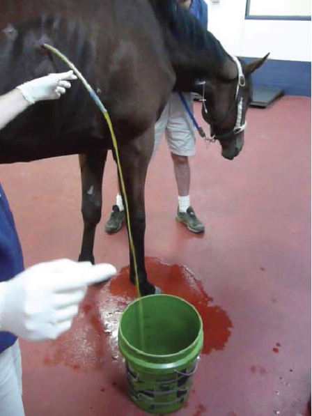
Between the sedation and Jen’s technique, Shiloh stayed still while Troy made an inch-long incision in the middle of the anesthetized area. He plunged a carmalt, a stainless-steel surgery pliers inside the incision, carefully rotating it back and forth pushing into the pleural space, the area outside the expanding lines was filling with fluid.
“I need to separate the muscle between these ribs, that’s why the lidocaine needle was so long.
When he was satisfied he retrieved the drain, a long white plastic tube eighteen inches long, and inserted it between the ribs.
Happy with the tube placement, Troy left the carmalts in place, holding both the tube and the carmalts with his left hand. This freed up his right to pull a large syringe from his pocket. He pushed air into the tube with the syringe to inflate a cuff round the end of the drain. The air-filled bubble would keep the surgical drain in place.
“Let’s see what we have,” he said, as he wiggled the drain, freeing up kinks and clogs. As if someone turned a spigot on, a rush of liquid came from the tube, surprising Troy who lost his grip on it. The tube’s end was flung around by the fast-moving fluid spraying onto the floor, flying everywhere, coating everything around it with the liquid from Shiloh's lungs. Thousands of infectious particles splattered hither and yon during the precious moments it took Troy to regain control of the tube.
Luckily Jen was on Shiloh’s other side and escaped the shower.
Shiloh started to struggle again. “Whoa, boy,” Troy said, as he wiped drops from his face and aimed the flow at the bucket.
“Stop, Shiloh!” Jen exclaimed sharply.
“Shh, softer, Jen, the sedative makes him sensitive to noise. Just tell him it's okay while you jiggle that twitch up and down.”
“That’s a lot of fluid, doc,” Jen exclaimed when she saw the bucket fill above the halfway point.
“Yeah, this should help a lot.”

This is the cytology from the trans-tracheal aspirate. The teeny tiny blue dots are bacteria. The large purple squiggly things are white cells called neutrophils and the biggest guys are macrophages. They are the large irregular balloon shaped cells, many of them have already ingested lots of bacteria. Photo is from Nottingham Veterinary School.
Just as Troy was relaxing, Shiloh suddenly twisted his head and ripped the twitch from Jen’s hand. It flew across the barn and clattered on the concrete. Shifting his feet, the horse kicked out and hit the bucket with his left rear, spilling the blood-tinged fluid onto the concrete where it rushed across the surface, spilling into cracks and crevices.
Then the horse turned his head toward Troy, who ducked and fell backward onto the wet concrete. His mouth hung open as he watched Shiloh rip the drain out with his teeth.
Troy was surprised, and shocked. “I think it’s time to stop for today,” he said. He wiped his sticky hands on his coveralls, grabbed the turned over bucket and picked up the discarded drain. “Make sure you clean the concrete with bleach, Jen. Use one-part bleach to water.”
“Why, are you worried?”
“I can’t go there, Jen. Let’s call it a precaution and just take this one step at a time.”
“Okay,” she said. She could see he was as shaken up as she was.
“We'll do the other side tomorrow.” He said as he gave the horse an antibiotic injection. “I'll leave these antibiotic tabs as well.”
“Dr. Osborn, could this fever be related to King’s illness?”
“Who?”
“He’s a Queensland Heeler I brought back from Australia and gave to Honey, Dr. Evan's assistant. He’s under Rory’s care, and he has a fever.”
“Well, that would be very unusual Jen, as it would mean it was a cross-species transfer. Make sure you bring it up with Rory, it could make us world famous.”
“Famous in a good or bad way, Dr. Osborne?”

This is photograph taken by Dr. Joe Pascoe at UCD showing the rib cage removed from the right side of a horse. The head is on the right, and the belly is on the left. Only a small section of normal lung tissue is seen in the upper left, it appears fluffy and light pink. The color darkens when infection sets in at first causing the consolidation seen in the middle. Finally at the lower right the lung lobe is completely obliterated with the purulent yellow exudate of infection.

or
or
Thank You!


Comments